
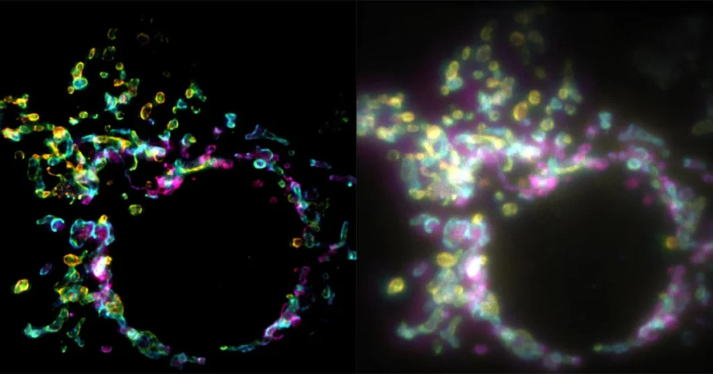

A breakthrough in optical engineering has arrived, promising to shift the paradigm of how biologists observe the microscopic world. Researchers at the École Polytechnique Fédérale de Lausanne (EPFL) have developed a novel fluorescence microscopy technique dubbed SPIFFI—Spatial Polarization-Induced Fluorescence Fluctuation Imaging. By enabling the capture of super-resolution images from a single exposure, this method overcomes the most significant bottleneck in modern cell biology: the trade-off between spatial resolution and temporal speed. The findings, published recently in the journal Nature, describe a technical framework that could render the traditional, multi-frame imaging processes that have long hampered the study of living cells obsolete.
The challenge of imaging living organisms at the nanoscale has historically been one of the most stubborn hurdles in the life sciences. While conventional light microscopy has provided invaluable insights into cellular structure, its utility is strictly bounded by the diffraction limit of light, which prevents researchers from clearly distinguishing objects smaller than approximately 200 to 300 nanometers. In the early 21st century, the development of super-resolution techniques—such as STED (Stimulated Emission Depletion) and PALM/STORM (Photoactivated Localization Microscopy/Stochastic Optical Reconstruction Microscopy)—allowed scientists to break this barrier, earning their developers the 2014 Nobel Prize in Chemistry.
However, these established techniques come with a heavy cost. To achieve high resolution, they typically require the acquisition of hundreds or even thousands of individual frames of the same specimen. Because cellular processes—such as mitochondrial fission, vesicle trafficking, and organelle fusion—occur on a millisecond timescale, the time required to collect these frames means the resulting image is often a blurry average of a moving target. In essence, the faster the cell moves, the less effective these super-resolution methods become.
The Mechanism Behind SPIFFI
SPIFFI represents a departure from this temporal-dependency model. Developed by a team led by Aleksandra Radenovic at EPFL’s Laboratory of Nanoscale Biology (LBEN), the technique leverages the physical properties of fluorescent light, specifically its polarization. Fluorescent molecules, or fluorophores, do not emit light in a uniform, isotropic manner; rather, they oscillate in directions dictated by their orientation and the constraints of their local environment.
The SPIFFI system functions by splitting incoming fluorescent light into four distinct polarization-sensitive channels. By performing a comparative analysis of these four channels, the system can reconstruct the spatial distribution of the molecules with a high degree of precision. This allows for the recovery of structural details that would otherwise be obscured by the diffraction limit in a standard widefield microscope.
Crucially, because all the information required to resolve the image is captured within a single exposure, the temporal resolution is limited only by the camera’s frame rate. This effectively enables the creation of high-definition, super-resolution video of living processes, a feat that was previously considered impractical or mathematically impossible with conventional stochastic super-resolution methods.
A Chronology of the Development
The development of SPIFFI was the result of a multi-year effort to refine optical instrumentation. The project began with the observation that the polarization data inherent in fluorescent emission was being largely discarded or treated as background noise in conventional imaging setups.
- Initial Concept (2023-2024): The research team hypothesized that by mapping the orientation of individual fluorescent dipoles to the structural context of the cell, they could bypass the need for temporal fluctuations.
- Prototype Design (2024): The team designed a custom optical pathway capable of splitting and detecting four polarization channels simultaneously.
- Validation Experiments (2025): The researchers utilized the SPIFFI setup to image mitochondrial membranes. The results showed a twofold increase in resolution compared to standard widefield imaging.
- Peer Review and Publication (2026): After extensive refinement, the findings were submitted to Nature, where they underwent rigorous scrutiny regarding the signal-to-noise ratio and the mathematical algorithms used to reconstruct the images. The paper was officially published in September 2026.
Quantitative Improvements and Capabilities
The performance metrics of SPIFFI offer a clear picture of why this technique is garnering attention. In its standalone configuration, SPIFFI achieves a spatial resolution of 160–170 nanometers. While this is already a significant improvement over the standard diffraction limit, the researchers found that it could be integrated with existing fluctuation-based post-processing methods. When these methods are combined, the resolution reaches approximately 80 nanometers.
This dual-mode capability is significant. For rapid, high-speed processes, researchers can rely on the single-frame SPIFFI output to track moving structures in real-time. For static or slower-moving structures where higher resolution is the priority, the integrated approach provides a powerful tool for detailed structural analysis.
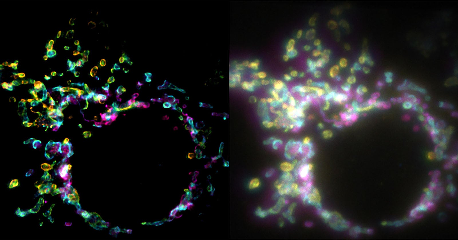
Wei Guo, the first author of the study and a PhD student at LBEN, notes the practical significance of this shift. "With previous techniques, taking many images would only result in one super-resolved frame," Guo stated. "With SPIFFI, every frame is super-resolved, meaning we can now produce super-resolution videos of live cells." This shift from static imaging to dynamic, high-speed, super-resolution cinematography allows researchers to observe events such as organelle division and vesicle transport with a level of clarity that was previously restricted to fixed, dead samples.
Practical Implementation and Accessibility
One of the most compelling aspects of the SPIFFI technology is its design philosophy regarding accessibility. Unlike some super-resolution platforms that require massive, dedicated, and incredibly expensive optical benches, the hardware components for SPIFFI were designed to be modular.
The researchers have successfully demonstrated that the SPIFFI optical hardware can be integrated into existing fluorescence microscopes currently in use in research laboratories worldwide. This "add-on" nature significantly lowers the barrier to entry for biological research teams. Instead of purchasing an entirely new system, labs can upgrade their existing imaging capabilities, provided they have the computational resources to process the polarization data.
The team is currently working on miniaturizing the optical components further. By making the SPIFFI module more compact, they hope to facilitate its adoption in a wider range of settings, including high-throughput screening environments where space and ease of use are at a premium.
Broader Implications for Cell Biology
The implications of SPIFFI extend far beyond the refinement of image resolution. By enabling high-throughput, multi-dimensional imaging, SPIFFI is poised to contribute to several critical areas of biological research:
- Neurobiology: Understanding the real-time dynamics of synaptic vesicles and axonal transport is crucial for studying neurodegenerative diseases like Alzheimer’s and Parkinson’s. SPIFFI’s ability to capture these processes without the "blur" of traditional methods could provide new insights into how these transport mechanisms fail.
- Cell Division and Oncology: By observing the high-speed mechanics of mitochondrial fission and fusion during cell division, researchers may uncover new markers for cancerous cells, which often exhibit abnormal organelle dynamics.
- Drug Discovery: High-throughput screening of drug candidates often relies on observing cellular responses to chemical stimuli. The ability to perform this screening at a super-resolved, dynamic level could lead to a more nuanced understanding of how pharmaceutical compounds interact with intracellular structures.
Expert Analysis and Future Directions
The scientific community has responded to the publication with optimism, noting that the combination of spatial and polarization-sensitive data is a logical next step in the evolution of light microscopy. Analysts at various imaging technology institutes have highlighted that while the technique is currently in the experimental stage, its ability to produce super-resolution video is a "game-changer" for those studying the kinetics of live cells.
"The real value here is the transition from ‘snapshot’ biology to ‘dynamic’ biology," said a representative from a leading biomedical imaging consortium. "By utilizing the polarization of light as a source of information rather than an obstacle, the researchers have opened a new window into the sub-cellular world."
Moving forward, the EPFL team plans to focus on the software side of the equation. While the hardware is ready for integration, the processing algorithms that interpret the polarization data can still be optimized for speed and accuracy. The team is also exploring the use of machine learning models to further refine the image reconstruction process, which could potentially push the resolution even further beyond the 80-nanometer threshold.
As the scientific community begins to adopt and adapt this technology, the expectation is that SPIFFI will become a standard component of the modern biological toolkit. By effectively solving the conflict between the need for speed and the need for precision, SPIFFI represents a vital advancement that aligns the capabilities of our optical tools with the frenetic, ever-changing reality of the living cell. The era of seeing the invisible in motion has, with this development, taken a definitive step forward.